This module has not been signed off by a clinician other than its author.
It implements the emergency management of chemical eye injury, the open globe, orbital compartment syndrome and traumatic hyphaema, drawn from RCEM Learning, the British Burn Association ocular burns guideline, and the lateral-canthotomy and hyphaema literature. Every quotation is verbatim from the source named beside it. But the implementation — which findings lead to which advice, and how the cross-injury cautions interact — has been checked only by the person who wrote it.
On a previous ResusDoc tool, an external reviewer found a serious error in a page that had already passed internal audit. Until an independent clinical review is complete, check anything you intend to act on against the source guidance and your local ophthalmology and eye-casualty pathway.
The reference
Chemical eye injury — irrigate before anything else
A chemical splash is the one eye emergency treated before history, examination or investigation. Alkalis (cement, plaster, oven and drain cleaner, ammonia) penetrate deeply and go on injuring; acids tend to coagulate and self-limit. A white, comfortable eye after an alkali burn is not reassurance — it is limbal ischaemia.
RCEM Learning & the British Burn Association guideline
- “Chemical eye injuries are true time-critical emergencies where immediate irrigation takes priority over history, examination or investigations.” (RCEM Learning)
- “Do not delay immediate irrigation of the eye for detailed assessment of patient or acquiring a particular irrigation fluid, regardless of delay in presentation.” (BBA UK CPG 2018)
- Anaesthetise, remove contact lenses and any particulate matter, and irrigate with a sterile isotonic solution (Hartmann's or 0.9% saline), an amphoteric solution (Diphoterine), or water. “Keep unaffected eye uppermost to avoid cross contamination.” (BBA)
- Target ocular pH 7.0–7.2; test the inferior fornix, and “repeat the pH test soon after the irrigation has ceased and again 30 minutes later” (BBA) — recheck every 5–10 minutes during irrigation and every 15 minutes for at least an hour once neutral, resuming if the pH drifts (RCEM Learning). Mean normal tear pH is 7.6.
- “A painless white eye may indicate severe limbal ischaemia rather than a minor injury.” Grade with Roper-Hall; Grade I can be discussed with ophthalmology, Grade II–IV need urgent referral. (RCEM Learning)
The Roper-Hall classification of ocular surface burns
Two things drive the prognosis: how clear the cornea is, and how much of the limbus — the ring of stem-cell-bearing vessels around the cornea — has been rendered ischaemic (blanched, non-perfused), measured in clock hours. The whiter and more comfortable the eye, the more limbus is dead.
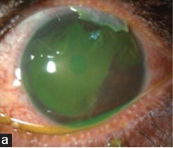
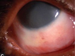
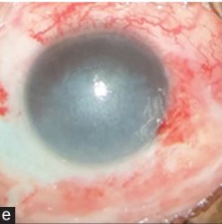
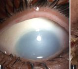
Clinical photographs: Iyer G, Srinivasan B, Agarwal S, Fogla R. E-PIX: An enhanced grading for acute ocular burns. Indian J Ophthalmol 2023;71(4):1401–1406 — panels cropped from Figs 1–2, reused under CC BY-NC-SA 4.0; this cropped adaptation is likewise offered under CC BY-NC-SA 4.0. Roper-Hall grade assigned here by the appearance of each eye (corneal clarity and limbal ischaemia), not by the source's own scale.
| Grade | Cornea | Limbal ischaemia | Prognosis |
|---|---|---|---|
| I | Corneal epithelial damage only | None | Good |
| II | Corneal haze, iris details visible | Less than ⅓ (under ~4 clock hours) | Good |
| III | Total epithelial loss, stromal haze obscuring iris details | ⅓ to ½ (~4–6 clock hours) | Guarded |
| IV | Cornea opaque, obscuring iris and pupil | More than ½ (over ~6 clock hours) | Poor |
Grading per Roper-Hall (1965), as tabulated by Gupta, Kalaivani & Tandon, Br J Ophthalmol 2011;95(2):194–8. The newer Dua classification extends this by grading limbal involvement in clock hours across six grades and predicts outcome better in the most severe burns; ophthalmology may use it in preference.
The open globe — shield it, and stop touching it
An open globe is a full-thickness wound of the cornea or sclera (Birmingham Eye Trauma Terminology: rupture from blunt force, or laceration — penetrating, perforating, or with a retained intraocular foreign body). Once you suspect one, the priority is to do no further harm: any pressure on the globe, including tonometry and an ultrasound probe, can extrude intraocular contents.
- Signs: a full-thickness wound, iris (uveal) prolapse, a shallow or flat anterior chamber, a teardrop or peaked pupil, a positive Seidel test, a deep lid laceration through the margin. A high-velocity mechanism (hammering metal, glass, power tools, blast) means an intraocular foreign body until proven otherwise.
- “Do not use eye patches in any circumstance, always use a protective eye shield which is rigid.” (RCEM Learning)
- “To avoid extrusion of intraocular contents, refrain from any unnecessary manipulation of the globe (including measuring intraocular pressure).” (RCEM Learning)
- Ultrasound is “contraindicated if suspecting open globe injury”; CT (1.5–2 mm axial and coronal cuts) is the investigation for a suspected foreign body. “A missed intraocular foreign body with ferrous component will lead to late vision loss due to siderosis.” (RCEM Learning)
- Keep nil by mouth for theatre; give analgesia and antiemetics to prevent Valsalva; start prophylactic systemic antibiotics against endophthalmitis (regimen per local protocol); check and update tetanus status; leave any protruding object in place. Urgent ophthalmology referral. (RCEM Learning; StatPearls)
- If intubation is needed for other injuries, succinylcholine raises intraocular pressure — a non-depolarising agent such as rocuronium is preferred, and ketamine is best avoided where possible (StatPearls). See the RSI planner — a trauma-team decision.
Orbital compartment syndrome & retrobulbar haemorrhage — the two-hour clock
Blood or oedema behind the globe raises pressure in a closed bony box, stretching and compressing the optic nerve. It is a clinical diagnosis — do not wait for the CT — and lateral canthotomy with inferior cantholysis is the sight-saving procedure. The evidence base is emergency-medicine and ophthalmology literature, not a UK national guideline.
- “Since OCS is a clinical diagnosis”, any patient with facial trauma or concern for retrobulbar haemorrhage “who presents with proptosis, decreased visual acuity, elevated intraocular pressures (IOP), restricted extraocular muscle movement, and/or afferent pupillary defect should be considered for emergent lateral orbital canthotomy with cantholysis”. (StatPearls)
- “Intraocular pressure measurements of 40 mmHg or higher indicate a soft threshold for intervention.” (StatPearls) — but the decision is the whole clinical picture, not one number, and IOP is not measured if an open globe is suspected.
- “Lateral canthotomy should be performed in the event of orbital compartment syndrome, ideally within 2 hours of presentation to avoid optic nerve ischaemia which will eventually lead to permanent loss of vision”, and “do not delay lateral canthotomy in known cases of retrobulbar hematoma” waiting for ophthalmology. (RCEM Learning)
- Contraindication: a ruptured globe — suggested by enophthalmos rather than proptosis, or confirmed on CT — is “a contraindication to the lateral orbital canthotomy” (StatPearls). Decompressing a ruptured globe extrudes its contents.
The procedure (RCEM Learning)
Infiltrate the lateral canthus with 1–2 mL of 1% lidocaine with adrenaline through a 25-gauge needle, directed away from the globe. Crimp the lateral corner of the lower lid with a haemostat to mark and thin it. Make a 1–2 cm incision from the lateral canthus, extending laterally. Identify the inferior crus of the lateral canthal tendon by blunt dissection, point the scissors inferiorly, strum it and cut until the lower lid is completely lax. Recheck the IOP; if still high, repeat on the superior crus. Note that a coexisting hyphaema can keep the pressure, the RAPD and the acuity abnormal even after a technically adequate cantholysis.
Full step-by-step: the lateral canthotomy & cantholysis procedure guide (ED Procedures) covers the equipment, the crush–cut–cantholyse sequence and the DOPS entrustment levels in detail.
Traumatic hyphaema — head up, shield, and think about sickle
A hyphaema is blood layering in the anterior chamber, usually from blunt trauma tearing iris or ciliary-body vessels. The acute dangers are a rise in intraocular pressure and a rebleed at day 2–5, which carries the worse prognosis.
- Grade by anterior-chamber filling: microhyphaema (scattered cells), I (<33%), II (33–50%), III (>50% but not total), IV (100%, “eight-ball”). Elevated IOP is seen in ~13.5% of grade I–II but ~52% of grade IV.
- “Hyphema treatment begins with elevating the head of the bed to at least 30 degrees”, so the blood layers below the visual axis; place a shield and keep it until the hyphaema resolves. (StatPearls)
- Measure IOP only once an open globe has been excluded; >21 mmHg is elevated and treated like acute glaucoma with ophthalmology input. Fluorescein for a corneal abrasion goes on before tonometry.
- “All patients should be cautioned against using non-steroidal anti-inflammatory agents, aspirin, and anticoagulants until the resolution of hyphema.” Control pain and nausea (vomiting spikes IOP and can restart bleeding). (StatPearls)
- Sickle cell disease or trait deserves special attention: the hypoxic anterior chamber induces sickling, blocking outflow and driving up pressure, and carbonic-anhydrase inhibitors (acetazolamide, dorzolamide) can worsen sickling. “Patients with a family history of sickle cell disease or trait or patients with uncertain status should be tested”, and their surgical thresholds are lower. (StatPearls)
- Antifibrinolytics (tranexamic / aminocaproic acid) are “no longer encouraged” for hyphaema — trials did not show reduced rebleeding. Rebleeding occurs in about 30%, at 2–5 days. (StatPearls) (This is the eye — it is not the trauma-wide TXA of NG39.)
- Outpatient management needs all of: grade ≤ II, normal IOP, no sickle disease/trait, no coagulopathy or bleeding diathesis, and the ability to attend daily ophthalmology review. Anyone failing these is admitted. All hyphaemas get an ophthalmology consultation.
Where NICE sits, and how these sources are tiered
NICE has essentially no guidance on the emergency management of ocular trauma. There is a Clinical Knowledge Summary on superficial corneal injury, and NG232 covers the head injury these patients so often carry, but neither addresses chemical burns, the open globe, orbital compartment syndrome or hyphaema. This is one of the few places on the trauma hub — like the penetrating-neck module — where the usual “NICE first” framing does not apply, and claiming NICE support here would be wrong.
So the sources are tiered honestly:
- RCEM Learning — the Royal College of Emergency Medicine's educational reference (CC-licensed), written for UK ED practice. Authoritative for our specialty, but educational material rather than a formal guideline.
- British Burn Association, Initial Management of Ocular Burns — a UK clinical practice guideline (2018). Its stated review date (June 2021) has passed; check the BBA site for a newer edition before relying on it for anything contentious.
- StatPearls (NCBI Bookshelf) — a US reference (CC BY-NC-ND), used for the canthotomy and hyphaema detail where no UK national guideline exists. The specific drug regimens it names are US practice; doses here defer to your local protocol.
- Roper-Hall classification — the standard ED grading of ocular chemical burns, cited via Gupta 2011.
Every quotation on this page is verbatim from the source named beside it. Where a statement is standard teaching rather than a cited guideline, it says so.
What this module deliberately does not do
- It is not the whole eye exam. Visual acuity, pupils and RAPD, lids and fornices (evert them), cornea and fluorescein/Seidel, anterior chamber, fundus and red reflex, and eye movements all still have to be done and documented — the mechanism has medicolegal weight.
- It does not cover the minor and the sub-acute: corneal abrasion, subtarsal foreign body, UV/arc-eye keratitis, subconjunctival haemorrhage, or the definitive management of an orbital “blow-out” fracture. Those are worked up once the four emergencies are excluded.
- It does not give a canthotomy competence. The steps are a reminder for a clinician already trained to do it, or working with telephone support from ophthalmology; it is not a substitute for training.
- It does not give drug doses. Antibiotic, analgesic and RSI doses are in the drug monographs and, for children, the paediatric tool.
Related
CT head & C-spine · Penetrating neck injury · Bleeding & anticoagulation reversal · Injury classifications · Drug monographs